ACOUSTIC NEUROMA
One of the possible causes for hearing troubles and development of deafness is acoustic neuroma. Аcoustic neuroma diagnostics includes audiometry and MRI scan. Аcoustic neuroma treatment consists of a wide range of conservative and surgical measures. More detailed information about this condition, as well as its treatment and diagnostics services you will find below.
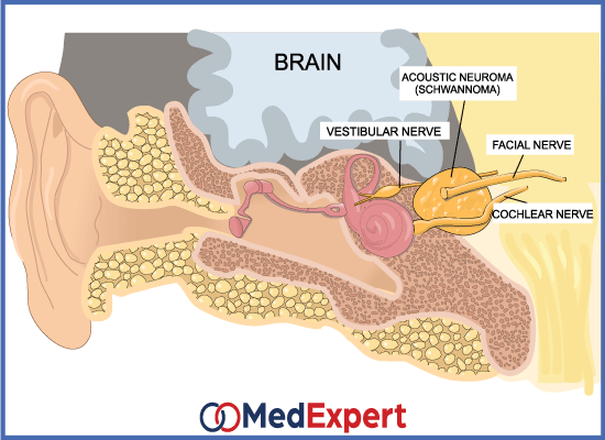
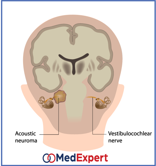
Acoustic neuroma is a rare, non-cancerous or benign and often slow growing tumour that develops on the main nerve leading from the inner ear to the brain. Pressure placed on branches of the main nerve directly influences balance and hearing as a result, acoustic neuroma can lead to hearing loss, ringing in ears and poor balance.
ARE ACOUSTIC NEUROMA TUMOURS DANGEROUS?
Acoustic neuromas which are also referred to as vestibular schwannomas or neurolemmomas normally develop slowly over many years; although they don’t invade the brain, they can apply pressure on it as they grow. The bigger tumours can exert pressure on the nerves controlling the muscles responsible for facial expression and sensation. Fatal results come across if the tumours grow large enough to press the brain stem or cerebellum.
SYMPTOMS OF ACOUSTIC NEUROMA
Symptoms experienced in acoustic neuroma are:
- Loss of balance
- Dizziness
- Numbness in the face
- Muscle weakness
- Ringing in the affected ear
ACOUSTIC NEUROMA TREATMENT OPTIONS
Acoustic neuroma has three major forms of treatment:
OBSERVATION
Due to the fact that acoustic neuroma form at a slow pace and are basically non-cancerous, treatment may not be required immediately. This still does not change the fact that doctors are likely to monitor the progress of the tumour via periodic MRI Scans. Other treatment measures may also be suggested in case there is continuous growth or side effects.
SURGERY
Sometimes, mere observation is not enough and a partial or complete removal of the tumor is needed. The approaches to the surgery are:
- Translabyrinthine: This procedure involves making a surgical cut behind the ear and the subsequent removal of the bone from behind the inner and outer ear. This procedure is only carried out when the tumor is beyond the minimum of 3cm. The con to this approach is that it can result in permanent loss of hearing.
- Retrosigmoid/Sub-Occipital: The skull is cut open close to the back of the head so as to expose the tumor. This option does not have any major drawbacks as it allows for tumors of any size to be removed while still maintaining patient’s hearing ability.
- Middle Fossa: The surgeon cuts out a part of the bone above the ear canal so as to allow access to the tumors within the internal auditory canal (the passage that connects the middle and inner ear to the brain). This procedure also retains the individual’s ability to hear.
Although the above surgical procedures are heavily in practice in modern times, there is a new technique that is less invasive which is known as endoscopic resection. This allows the surgeons to use a tiny camera to locate the acoustic neuroma by drilling a hole into the skull. Specially trained surgeons at high profile medical centers offer use this technique and it has recorded success rates that are similar to those of conventional surgery.
RADIATION THERAPY
This treatment is recommended in acoustic neuroma cases and it involves the use of high concentration of radiation to tumors while still trying to limit the damage to the other tissues. There are two radiation therapies available:
- Single fraction stereotactic radiosurgery: Every treatment session involves focusing tiny beams of radiation at the tumor.
- Multi-session fractionated stereotactic radiotherapy: The radiation treatment lasts for a couple of weeks and it is done on a daily basis.
Research shows that the multi-session therapy option of treatment has a better preservation rate for hearing when compared to the single fraction option.
ACOUSTIC NEUROMA DIAGNOSTICS SERVICES
- Physical examinations: The doctor enquires about the symptoms noticed and performs an evaluation on the health of the body. This is to attest that the symptoms are not as a result of other issues e.g. ear drums perforation.
- Hearing test (audiometry): The audiologist carries out this test to determine the patient’s ability to recognize sounds. The sounds are presented in different tones and the patient is asked to signify when the sounds are heard. The tones are set to determine what level the patient can hear at. In some cases, words can also be used.
- Scans: MRIs or CT scans are performed on the head so as to ascertain the presence of acoustic neuroma.