GLIOMA
It is a term used to describe any tumor that developed from the supported tissues (gluey) of the brain. This tissue is known as “glia” and it helps to keep the neurons in place and functioning properly.
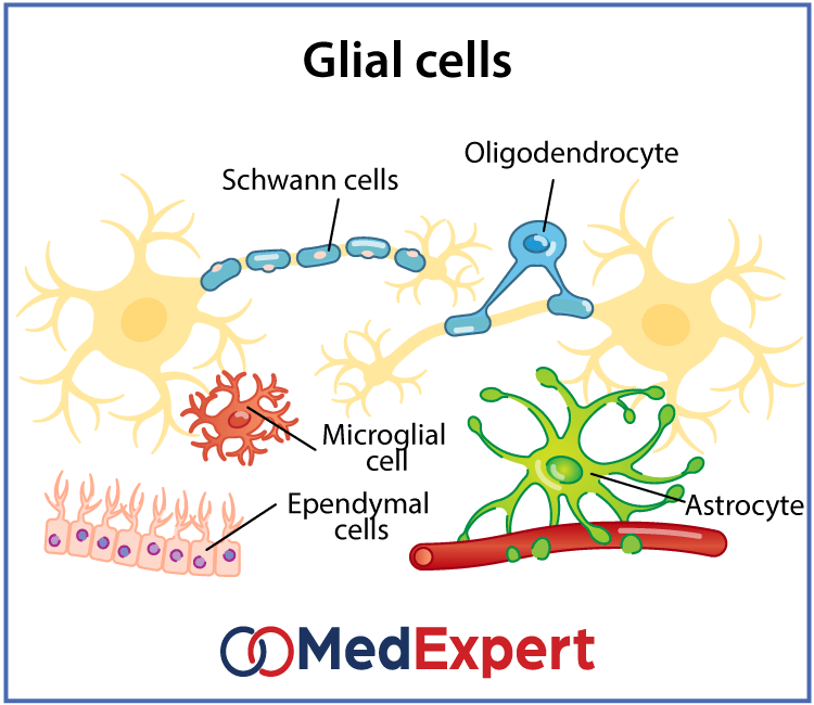
WHAT ARE THE THREE TYPES OF CELLS THAT CAUSE GLIOMA?
There are three types of normal glial cells that can cause glioma.
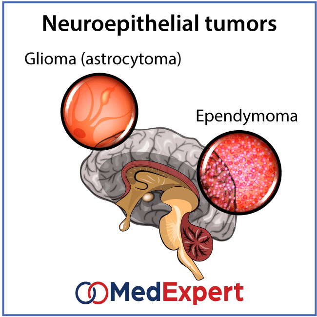
- Astrocyte: Produces astrocytomas and glioblastomas
- Oliogodendrocyte: Produces oligodendrogliomas
- Ependymal: Produces ependymomas
The tumors that show a mixture of these different cells are known as mixed gliomas. Other tumors such as optic nerve glioma and brain stem glioma are named after the locations they developed in and not the tissue type from which they originated.
SYMPTOMS OF GLIOMA
The signs and symptoms may vary depending on what type of tumor it is as well as the size, location and rate of growth. The signs and symptoms commonly experienced are:
- Headache
- Vomiting
- Nausea
- Confusion or a decline in brain function
- Memory loss
- Changes in personality and irritability
- Difficulty with balance
- Urinary incontinence
- Vision problems (blurred vision, double vision or loss of peripheral vision)
- Speech difficulties
- Seizures (especially in someone without a history of seizures)
TREATMENT OPTIONS AVAILABLE FOR GLIOMA
The type, size, grade and location of the tumor as well as your age, overall health and preferences will determine the treatment for glioma. Treatment for glioma may also require using drugs to minimize the symptoms and signs of your tumor. You may be prescribed steroids to help reduce swelling and relieve pressure on the affected areas of your brain. To help control seizures, anti-epileptic drugs may be used to control seizures.
Surgery
The first step in removing gliomas is to remove as much of the tumor as possible in all types of gliomas.
In cases where gliomas are small and easy to separate from surrounding brain tissue that’s healthy. This makes complete surgical removal possible. In other cases where your tumor is located near sensitive areas in your brain or can’t be removed from surrounding tissue, your surgeon will remove as much of the tumor as possible. Your signs and symptoms may be reduced even if a portion of the tumor is removed. In certain cases, the tissue samples that were removed by a surgeon will be analyzed by neuropathologists and the results will be reported while the surgery is underway. This will enable the surgeon on deciding how much tissue should be removed. A range of surgical technologies and techniques will be used to help your surgeon in protecting as much healthy brain tissue as possible while removing the tumor. The surgical technologies and techniques include:
- Computer-assisted brain surgery
- Awake brain surgery
- Intraoperative MRI
During awake brain surgery, you may be asked to move a limb or tell a story. This is to ensure that the areas of your brain controlling those functions are not compromised.
Intraoperative MRI
There are certain risks in removing a glioma through surgery such as infection and bleeding. Surgery poses other risks as well depending on which part of your brain is affected by the tumor. For example, a tumor near the nerves that connect to your eyes may carry a risk of vision loss if operated on.
Radiation therapy
After surgery, radiation therapy is normally required when treating glioma, especially high-grade gliomas. This therapy uses radiation uses high-energy beams like X-ray or protons to kill tumor cells.
The radiation treatment for glioma uses external beam radiation from a machine outside your body. Currently, there are several types of external beam radiation used and under study for glioma treatment. The timing and types of radiation therapy you may receive will depend on the type of glioma you have, the grade and other prognostic factors. Radiation therapy options include:
- Intensity-modulated radiation therapy (using computers to target the brain tum)
- Intensity-modulated radiation therapy (using computers to target the brain tumor)
- Proton beam therapy (using protons rather than X-rays as the source of radiation)
- Stereotactic radiation therapy (radiosurgery)
Gamma Knife targeting
In the traditional sense, radiosurgery is not surgery. Rather, it uses multiple radiation beams to give highly focused form of radiation treatment to destroy the tumor cells in small areas.
The individual beams are not particularly powerful. However, the point in which all the beams meet at the brain tumor receives a dose large enough to kill the tumor cells. To deliver radiation to treat the brain tumors, different types of technology are used in radiosurgery such as Gamma Knife or linear accelerator (LINAC). The side effects of radiotherapy will depend on the type and dose of radiation you receive. Some of the common side effects that will occurs during or immediately after radiosurgery include:
- Fatigue
- Headaches
- Scalp irritation
Chemotherapy
This treatment is used to kill tumor cells with drugs. These drugs can be taken in pill form (orally) or injected into your vein (intravenously).
This treatment is normally combined with radiation therapy to treat gliomas. The most often used drug in chemotherapy to treat glioma is temozolomide (temodar) which is consumed orally. The side effects of chemotherapy will vary depending on the type and dose of drugs received. The most common side effects of chemotherapy include:
- Nausea
- Vomiting
- Scalp irritation
- Headache
- Hair loss
- Fever
- Weakness
Some of these side effects can be managed with medication.
- Targeted drug therapy: This therapy focuses on certain abnormalities present in cancer cells. Targeted drug treatments can kill cancer cells by blocking these abnormalities.Bevacizumab (avastin) is a targeted drug therapy that is used to treat a type of brain cancer called glioblastoma. This drug is given intravenously. It stops the formation of new blood vessels, cutting off blood supply to a tumor and killing the tumor cells.
- Rehabilitation after treatment: As brain tumors develop in areas of the brain that control motor skills, vision, speech and thinking; rehabilitation may be required as part of your recovery. You may be referred to services that can help you by your doctor such as:
- Physical therapy: This can help you to regain lost motor skills or muscle strength
- Occupational therapy: This can help you return to your normal daily activities, including work (after a brain tumor or other illness)
- Speech therapy: If you have difficulty speaking, speech therapy with specialists in speech difficulties (speech pathologists) can help if you
- Tutoring for school-age children: This can help kids cope with the changes in their memory and thinking (after a brain tumor)
DIAGNOSING GLIOMA
- History taking and physical examination: Your doctor will ask you questions about your overall health, symptoms you have and family medical history, as those, who have a family history of cancers are assumed to be more predisposed to have the same condition.
- A neurological exam: Your doctor may check your vision, hearing, balance, coordination, strength and reflexes during a neurological exam. If there are problems in one of more of these parts may provide clues about the areas of your brain that could be affected by a brain tumor. A number of specialized MRI scan components may help your doctor evaluate the tumor and plan your treatment.
They include:
- Functional MRI
- Perfusion MRI
- Magnetic resonance spectroscopy
- CT scan
- PET scan
- Imaging tests: Magnetic resonance imaging (MRI) is most commonly used to help diagnose brain tumors. In certain cases, a contrast material may be injected in the vein of your arm during your MRI study to help show differences in brain tissue.
- Tests to find cancer in other parts of your body: Your doctor may recommend tests and procedures to determine the origin of the cancer to rule out other types of brain tumor that may have spread from other parts of the body. Gliomas are tumors that developed within the brain and are not a result of cancer that has spread from another part of the body.
- Collecting and testing a sample of abnormal tissue (biopsy): A needle biopsy before treatment or as part of an operation to remove the brain tumor depending on where the glioma is located in. A biopsy is the only definite way to diagnose a brain tumor and provide a prognosis to guide treatment decisions. A pathologist will be able to determine the grade of your brain tumor with the information provided.